각막 이물, Foreign body in the cornea(Corneal foreign body)
다음 정보를 참조(Please visit the following article) www.drleepediatrics.com-제1권 소아청소년 응급의료 제 2부 신체각계통 별 응급의료 제18장: 소아청소년 안과 질환 응급의료 참조 www.drleepediatrics.com-Volume 1 Emergency Medical Care for Children and Adolescents Part 2 Emergency Medical Care for Each System-Chapter 18: Pediatrics Eye Disease Emergency Medical Reference

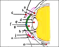
그림 52. 분흥색 선이 결막이다
a-모양체(섬모체), b-쉴렘관, c-위 눈꺼풀, d-홍채, e-수정체(렌즈), f-각막, g-전방, h-후방, i-아래 눈꺼풀, l-후방, n-결막, o-안구 하직근
Copyright ⓒ 2011 John Sangwon Lee, M.D., FAAP

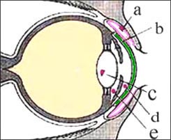
그림 53. 결막 이물과 안구 내 이물
분흥색 선이 결막이다
a-결막 이물, b-결막, c-수정체 내 이물, d-전방 내 이물, e-후방 내 이물
Copyright ⓒ 2011 John Sangwon Lee, M.D., FAAP
- 이물이 각막 표면에 살짝 붙어있든지 각막층 속 깊이 박혀 있을 수 있다. 이런 것을 각막 이물이라고 한다.
- 곤충, 먼지, 나무 부스러기 등 작은 것이 안구 속을 향해 힘없이 날아 들어오면 제일 먼저 속눈썹에 걸리거나 눈꺼풀을 꼭 감아 안구 속으로 더 이상 들어오지 못하거나, 일반적으로 안구로 들어온 후 결막 표면에 걸려 결막 표면 이물이 될 수 있다.
- 하지만 작은 쇠붙이나 날카로운 나무 부스러기, 마른 페인트 부스러기 등이 안구를 향해 힘세게 날아 들어올 때는 각막 표면 이물이 되거나 각막층 속 깊이 박히는 각막이물이 되든지, 또는 각막층을 지나 안구 속 더 깊이까지 깊이 들어갈 수 있다.
각막 이물의 증상 징후
- 각막의 이물의 크기, 이물의 종류, 각막의 표면에 이물이 있는지 각막층 속에 깊이 막혀 있는지, 합병증의 유무 등에 따라 증상 징후가 다르다.
- 각막 이물이 각막층 속에 깊이 박히면 눈이 상당히 아프고 눈물이 상당히 많이 난다.
- 햇빛이나 전등 불빛이 각막 이물이 있는 눈에 비칠 때 눈이 부시고 그 눈을 잘 뜰 수 없으며 이물이 생긴 눈이 당장에 빨갛게 된다.
- 각막 이물의 크기와 각막의 어느 부위에 이물이 있는지에 따라 시력 장애가 생길 수 있다.
각막 이물의 진단
- 병력 증상 징후와 진찰소견을 종합해서 진단할 수 있다.
- 각막 이물이 있다고 의심하면 눈을 절대로 비벼서는 안 된다.
- 거즈나 헝겊 조각으로 그 쪽 눈을 가리든지, 안대를 대고 안과로나 병원 응급실로 속히 데리고 가야 한다.
- 각막 표면에 살짝 걸친 이물은 자연히 빠져 나올 수 있다.
- 그렇지만 각막 표면에 살짝 묻힌 이물도 각막층 표면에 상처를 입힐 수 있다.
- 이물이 각막층 표면에 살짝 묻히든지 각막층 깊숙이 박혀있든지, 그 외 모든 종류의 각막 이물은 안과에서 치료 받아야 한다.
- 각막의 이물의 증상 징후와 각막 찰과상의 증상 징후가 서로 비슷할 때가 많다.
- 따라서 이 두 병을 감별 진단해서 치료해야 한다.
각막 이물의 치료
- 각막 이물이 있으면 안과에서 신속히 치료받아야 한다.
- 각막 이물이 각막 표면에 살짝 묻혀 있을 때도 안과에서 진단 치료를 받아야 한다.
- 일반적으로 안과에서는 각막 표면 이물은 쉽게 꺼낼 수 있다.
- 그렇지만 각막층 깊숙이 박힌 이물은 병원에서 전신 마취 하에서 꺼내는 것이 보통이다.
- 각막 이물로 생긴 각막 상처도 동시 치료받아야 한다.
- 각막 이물을 신속히 치료해야 한다.
- 각막 이물을 신속히 치료해 주지 않았을 때는 박테리아 감염이 각막 이물로 생긴 각막 상처에 생겨 각막염이나 안구염이 생길 수 있다.
- 각막염으로 시력 장애가 생길 수 있고, 심할 때는 실명할 수 있다.
- 따라서 각막 이물은 조기 진단 치료해야 한다.
- 결막 이물, 눈의 이물, 안구 속에 들어간 이물, 각막 외상
Foreign body in the cornea (Corneal foreign body)
Please visit the following article www.drleepediatrics.com-Volume I. Emergency Medical Care for Children and Adolescents Part II. Volume 1 Emergency Medical Care for Children and Adolescents Part 2 Emergency Medical Care for Each System-Chapter 18: Pediatrics Eye Disease Emergency Medical Reference

Figure 52. The purple line is the conjunctiva a-ciliary body (ciliary body), b-Schlem’s canal, c-upper eyelid, d-iris, e-lens (lens), f-cornea, g-anterior, h-posterior, i-lower eyelid, l-posterior, n – conjunctiva, o – inferior rectus muscle Copyright ⓒ 2011 John Sangwon Lee, M.D., FAAP

Figure 53. Conjunctival foreign body and intraocular foreign body The purple line is the conjunctiva a-conjunctival foreign body, b-conjunctival foreign body, c-intraocular foreign body, d-anterior intraocular foreign body, e-posterior intraocular foreign body Copyright ⓒ 2011 John Sangwon Lee, M.D., FAAP
• The foreign body may be slightly attached to the surface of the cornea or may be deeply embedded in the corneal layer. This is called a corneal foreign body.
• When a small object such as an insect, dust, or wood shavings flies into the eye without force, the first thing to get caught is the eyelashes or close the eyelids so that it cannot enter the eye anymore, or usually after entering the eye, it gets caught on the surface of the conjunctiva. can be a foreign object
• However, when small metal objects, sharp wood chips, or dry paint chips, etc. are blown into the eyeball forcefully, they become a foreign object on the surface of the cornea, become a foreign object that penetrates deeply into the corneal layer, or passes through the corneal layer and enters deeper into the eye.
Symptoms of a corneal foreign body
• Symptoms differ depending on the size of the foreign object in the cornea, the type of foreign object, whether there is a foreign object on the surface of the cornea or deeply blocked in the corneal layer, and whether there are complications.
• When a corneal foreign body is deeply embedded in the corneal layer, the eye hurts considerably and tears flow considerably.
• When sunlight or electric light shines on the eye with a corneal foreign body, the eyes become dazzled and unable to open, and the foreign body eye immediately turns red.
• Depending on the size of the corneal foreign body and where the foreign body is located, vision impairment may occur.
Diagnosis of a corneal foreign body
• The diagnosis can be made by combining the medical history, symptoms, signs, and examination findings.
• Never rub your eyes if you suspect you have a corneal foreign body.
• Cover the eye with gauze or a piece of cloth, or wear an eye patch and take it to the eye doctor or hospital emergency room promptly.
• A foreign body slightly over the corneal surface can come out naturally.
• However, even a foreign object lightly deposited on the surface of the cornea can injure the surface of the corneal layer.
• Whether the foreign body is lightly buried on the surface of the corneal layer or deeply embedded in the corneal layer, all other types of corneal foreign body should be treated by an ophthalmologist.
• The symptoms of corneal foreign body and corneal abrasions are often similar.
• Therefore, these two diseases should be differentially diagnosed and treated.
Treatment of corneal foreign bodies
• If there is a corneal foreign body, it should be treated promptly by an ophthalmologist.
• Even when a corneal foreign body is lightly buried on the corneal surface, it is necessary to seek diagnostic treatment from an ophthalmologist.
• In general, the corneal surface foreign body can be easily removed in ophthalmology.
• However, foreign bodies embedded deep in the corneal layer are usually removed at the hospital under general anesthesia.
• Corneal wounds caused by corneal foreign bodies should also be treated at the same time.
• The corneal foreign body should be treated promptly.
• If the corneal foreign body is not treated promptly, a bacterial infection can develop in the corneal wound caused by the corneal foreign body, resulting in keratitis or ophthalmitis.
• Keratitis can impair your vision and, in severe cases,
cause blindness.
• Therefore, corneal foreign bodies should be diagnosed and treated early.
• Conjunctival foreign body, foreign body in the eye, foreign body in the eyeball, corneal trauma
출처 및 참조 문헌 Sources and references
- NelsonTextbook of Pediatrics 22ND Ed
- The Harriet Lane Handbook 22ND Ed
- Growth and development of the children
- Red Book 32nd Ed 2021-2024
- Neonatal Resuscitation, American Academy of Pediatrics
- www.drleepediatrics.com 제1권 소아청소년 응급 의료
- www.drleepediatrics.com 제2권 소아청소년 예방
- www.drleepediatrics.com 제3권 소아청소년 성장 발육 육아
- www.drleepediatrics.com 제4권 모유,모유수유, 이유
- www.drleepediatrics.com 제5권 인공영양, 우유, 이유식, 비타민, 미네랄, 단백질, 탄수화물, 지방
- www.drleepediatrics.com 제6권 신생아 성장 발육 육아 질병
- www.drleepediatrics.com제7권 소아청소년 감염병
- www.drleepediatrics.com제8권 소아청소년 호흡기 질환
- www.drleepediatrics.com제9권 소아청소년 소화기 질환
- www.drleepediatrics.com제10권. 소아청소년 신장 비뇨 생식기 질환
- www.drleepediatrics.com제11권. 소아청소년 심장 혈관계 질환
- www.drleepediatrics.com제12권. 소아청소년 신경 정신 질환, 행동 수면 문제
- www.drleepediatrics.com제13권. 소아청소년 혈액, 림프, 종양 질환
- www.drleepediatrics.com제14권. 소아청소년 내분비, 유전, 염색체, 대사, 희귀병
- www.drleepediatrics.com제15권. 소아청소년 알레르기, 자가 면역질환
- www.drleepediatrics.com제16권. 소아청소년 정형외과 질환
- www.drleepediatrics.com제17권. 소아청소년 피부 질환
- www.drleepediatrics.com제18권. 소아청소년 이비인후(귀 코 인두 후두) 질환
- www.drleepediatrics.com제19권. 소아청소년 안과 (눈)질환
- www.drleepediatrics.com 제20권 소아청소년 이 (치아)질환
- www.drleepediatrics.com 제21권 소아청소년 가정 학교 간호
- www.drleepediatrics.com 제22권 아들 딸 이렇게 사랑해 키우세요
- www.drleepediatrics.com 제23권 사춘기 아이들의 성장 발육 질병
- www.drleepediatrics.com 제24권 소아청소년 성교육
- www.drleepediatrics.com 제25권 임신, 분만, 출산, 신생아 돌보기
- Red book 29th-31st edition 2021
- Nelson Text Book of Pediatrics 19th- 21st Edition
- The Johns Hopkins Hospital, The Harriet Lane Handbook, 22nd edition
- 응급환자관리 정담미디어
- Pediatric Nutritional Handbook American Academy of Pediatrics
- 소아가정간호백과–부모도 반의사가 되어야 한다, 이상원 저
- The pregnancy Bible. By Joan stone, MD. Keith Eddleman, MD
- Neonatology Jeffrey J. Pomerance, C. Joan Richardson
- Preparation for Birth. Beverly Savage and Dianna Smith
- 임신에서 신생아 돌보기까지. 이상원
- Breastfeeding. by Ruth Lawrence and Robert Lawrence
- Sources and references on Growth, Development, Cares, and Diseases of Newborn Infants
- Emergency Medical Service for Children, By Ross Lab. May 1989. p.10
- Emergency care, Harvey Grant and Robert Murray
- Emergency Care Transportation of Sick and Injured American Academy of Orthopaedic Surgeons
- Emergency Pediatrics A Guide to Ambulatory Care, Roger M. Barkin, Peter Rosen
- Quick Reference To Pediatric Emergencies, Delmer J. Pascoe, M.D., Moses Grossman, M.D. with 26 contributors
- Neonatal resuscitation Ameican academy of pediatrics
- Pediatric Nutritional Handbook American Academy of Pediatrics
- Pediatric Resuscitation Pediatric Clinics of North America, Stephen M. Schexnayder, M.D.
-
Pediatric Critical Care, Pediatric Clinics of North America, James P. Orlowski, M.D.
-
Preparation for Birth. Beverly Savage and Dianna Smith
-
Infectious disease of children, Saul Krugman, Samuel L Katz, Ann A.
-
Emergency Care Transportation of Sick and Injured American Academy of Orthopaedic Surgeons
-
Emergency Pediatrics A Guide to Ambulatory Care, Roger M. Barkin, Peter Rosen
-
Gray’s Anatomy
-
제19권 소아청소년 안과 질환 참조문헌 및 출처
- Habilitation of The handicapped Child, The Pediatric Clinics of North America, Robert H Haslam, MD.,
-
Pediatric Ophthalmology, The Pediatric Clinics of North America, Leonard B. Nelson, M.D.
-
Pediatric Ophthalmology, The Pediatric Clinics of North America, Lois J. Martyn, M.D.
-
Pediatric Ophthalmology, Edited by Robison D. Harley, M.D.
-
The Pediatric Clinics of North America, David Tunkel, MD., Kenneth MD Grundfast, MD
-
Atlas Pediatric Physical Diagnosis Frank A Oski
Copyright ⓒ 2014 John Sangwon Lee, MD., FAAP
“부모도 반의사가 되어야 한다”-내용은 여러분들의 의사로부터 얻은 정보와 진료를 대신할 수 없습니다.
“The information contained in this publication should not be used as a substitute for the medical care and advice of your doctor. There may be variations in treatment that your doctor may recommend based on individual facts and circumstances.
“Parental education is the best medicine.