결막 이물, Foreign bodies in the conjunctiva
다음 정보를 참조(Please visit the following article) www.drleepediatrics.com-제1권 소아청소년 응급의료 제 2부 신체각계통 별 응급의료 .제18장 : 소아청소년 안과 질환 응급의료 참조 www.drleepediatrics.com-Volume 1 Emergency Medical Care for Children and Adolescents Part 2 Emergency Medical Care for Each System-Chapter 18: Pediatrics Eye Disease Emergency Medical Reference
- 눈에 이물이 들어가 있는 것을 눈의 이물이라고 한다.
- 결막, 각막 속으로 들어온 연기나 먼지 등 작은 이물도 일종의 눈의 이물이다.
- 교통사고가 날 때나, 운동을 할 때, 또는 장난치다가 나무 부스러기, 모래알, 곤충, 쇠붙이 등이 결막, 각막, 또는 안구 속으로 세게 들어와 눈에 이물이 생길 수 있다.
- 이물이 들어 있는 눈의 부분에 따라 결막 이물, 각막 이물, 안구 내 이물 등으로 분류된다.
- 더 자세한 눈의 이물에 관해 다음 각 항을 참조.
- 결막 이물, 각막 이물, 안구 속에 들어간 이물, 각막 외상 (Corneal trauma)
- 결막층의 표면이나 결막층 속에 있는 이물을 결막 이물이라고 한다.
- 모래, 먼지, 곤충, 나뭇조각 등 이물이 결막 표면에 들어와 있거나 결막에 묻힐 수 있다. 흡인구 사진 참조.
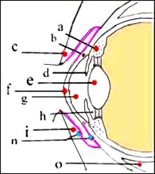
그림 48. 부분이 결막이다
a-모양체(섬모체), b-쉴렘 관, c-위 눈꺼풀, d-홍채, e-수정체(렌즈), f-각막, g-전방, h-후방, i-아래 눈꺼풀, l-후방, n-결막, o-안구 하직근
Copyright ⓒ 2011 John Sangwon Lee, M.D., FAAP
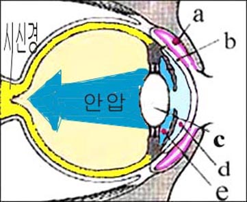
그림 49. 결막 이물과 안구 내 이물
분흥색 부분이 결막이다
a-결막 이물, b-결막, c-수정체 내 이물, d-전방내 이물, e-후방내 이물
Copyright ⓒ 2011 John Sangwon Lee, M.D., FAAP
그림 50. 하 결막 이물
Copyright ⓒ 2011 John Sangwon Lee, M.D., FAAP
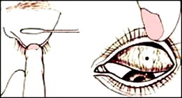
그림 51. 상 결막 이물
결막충의 이물과 안구 내 이물
Copyright ⓒ 2011 John Sangwon Lee, M.D., FAAP
결막 이물의 증상 징후
- 이물이 결막 표면에 있는지, 결막층 속에 묻혀 있는지, 이물의 종류와 환아의 나이 등에 따라 결막 이물의 증상 징후가 다르다.
- 연기나 미세한 먼지 등이 결막의 표면에 있을 때에는 눈물이 금방 많이 나고 그 눈물로 결막 표면 이물이 자연적으로 씻겨 눈 밖으로 나오는 것이 보통이다.
- 그렇지만 결막 표면 큰 이물이나 결막층 속에 꼭 박힌 결막 이물이 있을 때에는 여러 가지 증상 징후가 생길 수 있다.
- 눈에 어떤 이상이 있다고 말로 잘 표현할 줄 모르는 영유아들의 결막 표면에 이물이 있을 때나 이물이 결막층 속에 묻혀 있을 때는 이물이 있는 눈을 정상적으로 뜨기 싫어한다. 그리고 눈을 비비기도 하고 울기도 한다.
- 결막 표면 이물이 있는 눈을 비빌 때 결막이 더 충혈 되어 눈이 더 빨갛게 될 수 있다.
- 말로 표현을 할 수 있는 아이의 결막의 표면에 이물이 들어갔을 때는 눈에 무엇이 들어갔다고 말하기도 하고 눈이 아프다고 말하든지, 또는 눈이 꺼칠꺼칠하다고 표현 할 수 있다.
결막 이물의 진단
- 병력 증상 징후와 진찰소견 등을 종합해 진단한다.
- 결막 표면에 있는 결막 이물의 대부분은 육안으로 보고 쉽게 진단할 수 있다.
- 결막 층 속에 박힌 이물은 안과 전문의의 진단 치료를 받아야 한다.
결막 이물의 치료
- 증상 징후가 심하지 않고, 결막층 속에 깊숙이 박히지 않고 결막 표면에 있고 눈으로 쉽게 볼 수 있는 이물은 눈물로 씻겨 자연적으로 나올 수도 있고, 그렇게 자연히 나오지 않는 결막 표면 이물은 부모가 집에서 잘 처치하면 이물을 쉽게 꺼낼 수 있다.
- 자녀가 부모의 말을 잘 듣고 결막 표면에 있는 이물을 꺼내도 좋다고 협조적이고 결막 표면 이물을 꺼내고 좋다고 허락할 때는 부모가 집에서 다음 방법으로 결막 이물을 꺼낼 수 있다.
결막 표면 이물 꺼내는 방법
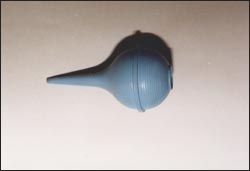
사진. 흡인구(빨기 기구)
Copyright ⓒ 2011 John Sangwon Lee, M.D., FAAP
① 눈을 더 이상 비비지 않게 주의를 준다. 결막 표면 이물을 꺼내주기 전에 그 아이에게 이물을 꺼내는 이유와 방법에 대해서 간단히 설명 한다.
② 결막 표면 이물을 꺼내주기 전에 꺼내줄 사람은 비눗물로 손을 깨끗이 씻는다.
③ 화아가 부모의 무릎 위에 아이의 머리를 대고 눕히든지 바닥에 눕힌다.
④ 결막 표면 이물이 있는 눈을 크게 떴다 감았다 한두 번하게 한다. 눈을 떴다 감았다 할 때 결막 표면 이물이 결막 표면의 한 부위에서 다른 부위로 이동하는지 관찰한다.
자녀가 눈을 떴다 감았다 할 수 없을 때는 처치자의 한 손가락 끝으로 아래눈꺼풀을 조심스럽게 아래로 까서 벌리고 그 눈의 결막 표면에 전등불을 비춰가면서 결막 표면에 이물이 있나 찾아본다.
⑤ 한 손가락 끝으로 아래 눈꺼풀을 아래로 까 젖혀 뒤집고 전등불을 결막에 비춰 결막 표면 어느 부위에 이물이 있나 찾아본다. (그림 50 참조)
이물이 결막층 속에 깊이 박혀 있지 않고 결막 표면에 있을 때는 이물을 면봉 끝으로 살짝 묻혀 꺼내든지 흡인구 속 멸균수나 생리식염수로 결막 표면 이물을 씻어낼 수 있다. 때로는 컵에 넣은 수돗물로 결막 표면 이물을 씻어낼 수 있다. 또는 흡인구가 없으면 깨끗이 씻은 손으로 깨끗한 물을 떠서 결막 이물을 씻어낼 수 있다.
⑥ 이물이 아래 눈꺼풀의 안쪽 결막 표면이나 결막층 속에 깊이 박혀 있지 않을 때는 이물이 위 눈꺼풀의 안쪽 결막 표면에 있을 가능성이 많다.
이때는 그림 51과 같이 윗 눈꺼풀을 뒤집고 윗 눈꺼풀 안쪽 결막 표면에 이물이 있나 찾아본다. 이물이 있으면 위에서와 같은 요령으로 꺼낸다.
⑦ 결막 표면 이물을 꺼내도 괜찮다고 허락 받고 부모가 아이의 결막 이물을 꺼내는 동안 갑자기 협조하지 않을 수 있다. 부모가 면봉 등으로 결막 이물을 꺼낼 때 그 아이가 부모의 손에 잡은 면봉 등을 갑자기 쳐서 눈에 큰 상처를 입을 수 있다.
아이가 결막 표면 이물을 꺼내도 괜찮다고 허락을 했든 안 했든, 결막 표면 이물을 집에서 꺼낼 때는 아이의 머리와 두 손을 꼭 붙들고 안전하게 꺼내야 한다.
⑧ 이물이 결막 표면에 확실히 있지만 부모가 결막 표면 이물을 꺼내 줄 자신이 없거나, 아이가 이물을 꺼내는 데 전혀 협조하지 않을 때는 안과 전문의나 소아청소년과 전문의, 또는 병원 응급실에서 이물을 꺼내야 한다.
⑨ 아주 작은 먼지 등이 눈에 많이 들어가서 결막 표면에 많이 붙어 있을 때는 눈을 떴다 감았다 하면서 깨끗한 수돗물을 손으로 떠서 눈을 충분히 씻어주면 작은 먼지는 자연히 나오게 된다.
이때 깨끗한 수돗물 대신 생리식염수(0.9% 식염수)를 써도 된다.
수돗물을 손으로 떠서 결막을 씻어주는 대신 흐르는 수돗물이 눈의 결막에 직접 흐르게 해서 결막 표면 이물을 씻어 낼 수도 있고, 흡인구에 깨끗한 수돗물이나 생리 식염수를 넣어서 결막을 씻어 주어도 된다.
⑩ 결막에 깊이 박힌 이물은 병원에서 빼야 한다.
Foreign bodies in the conjunctiva
Please visit the following article www.drleepediatrics.com-Volume 1 Emergency Medical Care for Children and Adolescents Part 2 Emergency Medical Care for Each System. See Chapter 18: Emergency Medical Care for Pediatric Eye Diseases www.drleepediatrics.com -Volume 1 Emergency Medical Care for Children and Adolescents Part 2 Emergency Medical Care for Each System-Chapter 18: Pediatrics Eye Disease Emergency Medical Reference
• A foreign body in the eye is called a foreign body.
• Small foreign objects such as smoke or dust that have entered the conjunctiva or cornea are also a type of foreign object in the eye.
• In the event of a traffic accident, exercise, or play, wood shavings, sand grains, insects, or metal may enter the conjunctiva, cornea, or eyeball hard and cause a foreign body in the eye. • According to the part of the eye where the foreign body is contained, it is classified into a conjunctival foreign body, a corneal foreign body, and an intraocular foreign body.
• For more details on foreign bodies in the eye, see each of the following sections.
• Conjunctival foreign body, corneal foreign body, intraocular foreign body, corneal trauma
• Foreign substances on the surface of the conjunctival layer or in the conjunctival layer are called conjunctival foreign substances.
• Foreign objects such as sand, dust, insects, and wood chips may enter the surface of the conjunctiva or may be deposited on the conjunctiva. See picture of suction port.
Figure 48. Partial conjunctiva a-ciliary body (ciliary body), b-Schlem’s canal, c-upper eyelid, d-iris, e-lens (lens), f-cornea, g-anterior, h-posterior, i-lower eyelid, l-posterior, n – conjunctiva, o – inferior rectus muscle Copyright ⓒ 2011 John Sangwon Lee, M.D., FAAP
Figure 49. Conjunctival foreign body and intraocular foreign body The purple part is the conjunctiva a-Conjunctival foreign body, b-Conjunctival foreign body, c-Intraocular foreign body, d-Intra-anterior foreign body, e-Posterior intraocular foreign body Copyright ⓒ 2011 John Sangwon Lee, M.D., FAAP
Figure 50. Inferior conjunctival foreign body Copyright ⓒ 2011 John Sangwon Lee, M.D., FAAP
Figure 51. Upper conjunctival foreign body Conjunctival foreign body and intraocular foreign body Copyright ⓒ 2011 John Sangwon Lee, M.D., FAAP
Signs of symptoms of a conjunctival foreign body
• Symptoms of conjunctival foreign body differ depending on whether the foreign body is on the surface of the conjunctiva or is buried in the conjunctival layer, the type of foreign body and the age of the child.
• When smoke or fine dust is on the surface of the conjunctiva, tears come out quickly, and it is normal for the conjunctiva surface foreign substances to be washed out naturally with the tears.
• However, when there is a large foreign body on the surface of the conjunctiva or a conjunctival foreign body embedded in the conjunctival layer, various symptoms may occur.
• When there is a foreign object on the surface of the conjunctival conjunctiva or when a foreign object is buried in the conjunctival layer, infants and young children who do not know how to verbally express that there is something wrong with their eyes do not like to open their eyes normally. And I rub my eyes and cry.
• Rubbing an eye with a conjunctival surface foreign body can make the conjunctiva more red, making the eye more red.
• When a foreign object enters the surface of a child’s conjunctiva, which can be expressed verbally, it can be said that something has entered the eye, that the eye is painful, or that the eye is rough.
Diagnosis of conjunctival foreign body
• Diagnosis is made by synthesizing medical history, symptoms, signs, and examination findings.
• Most of the conjunctival foreign bodies on the conjunctival surface can be easily diagnosed with the naked eye. • Foreign bodies embedded in the conjunctival layer should be diagnosed and treated by an ophthalmologist. treatment of conjunctival foreign body
• Foreign objects that are not symptomatic, are not deeply embedded in the conjunctival layer, are on the surface of the conjunctiva and are easily visible to the eye, may come out naturally by washing with tears. can be taken out easily.
• When the child listens to the parents and cooperates to remove the foreign body from the conjunctival surface and agrees to take it out, the parent can remove the foreign body from the conjunctival surface in the following way at home.
How to remove a foreign body from the conjunctival surface
Photo. Suction port (sucking device) Copyright ⓒ 2011 John Sangwon Lee, M.D., FAAP
① Be careful not to rub your eyes any more. Before removing the conjunctival surface foreign body, briefly explain the reason and method of removing the foreign body to the child.
② Before removing the conjunctival surface, the person who will take it out should wash their hands thoroughly with soapy water.
③ Hwa-ah lays the child’s head on the parent’s lap or lays it on the floor.
④ Open and close the eyes with foreign objects on the surface of the conjunctiva once or twice. Observe whether foreign objects on the surface of the conjunctiva move from one part of the surface of the conjunctiva to another when the eyes are opened and closed.
If your child is unable to open or close his/her eyes, carefully open the lower eyelid downwards with the tip of one finger of the caregiver and shine a light on the surface of the conjunctiva to check for foreign objects on the surface of the conjunctiva.
⑤ With the tip of one finger, flip the lower eyelid down and turn it over and look for any foreign objects on the surface of the conjunctiva by shining a light on the conjunctiva. (See Figure 50) If the foreign material is not deeply embedded in the conjunctival layer and is on the surface of the conjunctiva, gently apply the foreign material with the tip of a cotton swab and take it out, or wash the surface of the conjunctiva with sterile water or physiological saline in the suction port.
Occasionally, tap water placed in a cup can flush out the conjunctival surface. Alternatively, if there is no suction hole, you can wash the conjunctival foreign material by scooping clean water with clean hands.
⑥ When the foreign body is not deeply embedded in the inner conjunctival surface or the conjunctival layer of the lower eyelid, there is a high probability that the foreign object is on the inner conjunctival surface of the upper eyelid. In this case, as shown in Figure 51, turn the upper eyelid over and look for foreign objects on the surface of the conjunctiva inside the upper eyelid.
If there is a foreign object, take it out in the same way as above.
⑦ The parent may suddenly not cooperate while the child’s conjunctival foreign body is removed after being allowed to remove the conjunctival surface foreign body. When a parent removes a conjunctival foreign body with a cotton swab, etc., the child may suddenly hit the cotton swab, etc. held in her parent’s hand, causing a serious injury to her eye. Whether or not the child has given permission to remove the conjunctival surface object, when removing the conjunctival surface object from the house, hold the child’s head and both hands tightly and take it out safely.
⑧ When a foreign object is clearly on the surface of the conjunctiva, but the parent is not confident to remove the foreign object from the conjunctival surface, or the child does not cooperate at all in removing the foreign object, an ophthalmologist, a pediatric specialist, or a hospital emergency room should remove the foreign object.
⑨ When a lot of very small dust enters the eye and is attached to the surface of the conjunctiva, open and close the eye and wash the eye sufficiently with clean tap water by hand, and the small dust will come out naturally. In this case, you can use physiological saline (0.9% saline) instead of clean tap water.
Instead of washing the conjunctiva by scooping out tap water by hand, flowing tap water can flow directly to the conjunctiva of the eye to wash away foreign substances on the conjunctival surface, or you can wash the conjunctiva by adding clean tap water or physiological saline to the suction port. ⑩ Foreign objects deeply embedded in the conjunctiva should be removed from the hospital.
출처 및 참조 문헌 Sources and references
- NelsonTextbook of Pediatrics 22ND Ed
- The Harriet Lane Handbook 22ND Ed
- Growth and development of the children
- Red Book 32nd Ed 2021-2024
- Neonatal Resuscitation, American Academy of Pediatrics
- 눈의 이물
- 각막 이물
- 안구 속에 들어간 이물
- 각막 외상
- www.drleepediatrics.com 제1권 소아청소년 응급 의료
- www.drleepediatrics.com 제2권 소아청소년 예방
- www.drleepediatrics.com 제3권 소아청소년 성장 발육 육아
- www.drleepediatrics.com 제4권 모유,모유수유, 이유
- www.drleepediatrics.com 제5권 인공영양, 우유, 이유식, 비타민, 미네랄, 단백질, 탄수화물, 지방
- www.drleepediatrics.com 제6권 신생아 성장 발육 육아 질병
- www.drleepediatrics.com제7권 소아청소년 감염병
- www.drleepediatrics.com제8권 소아청소년 호흡기 질환
- www.drleepediatrics.com제9권 소아청소년 소화기 질환
- www.drleepediatrics.com제10권. 소아청소년 신장 비뇨 생식기 질환
- www.drleepediatrics.com제11권. 소아청소년 심장 혈관계 질환
- www.drleepediatrics.com제12권. 소아청소년 신경 정신 질환, 행동 수면 문제
- www.drleepediatrics.com제13권. 소아청소년 혈액, 림프, 종양 질환
- www.drleepediatrics.com제14권. 소아청소년 내분비, 유전, 염색체, 대사, 희귀병
- www.drleepediatrics.com제15권. 소아청소년 알레르기, 자가 면역질환
- www.drleepediatrics.com제16권. 소아청소년 정형외과 질환
- www.drleepediatrics.com제17권. 소아청소년 피부 질환
- www.drleepediatrics.com제18권. 소아청소년 이비인후(귀 코 인두 후두) 질환
- www.drleepediatrics.com제19권. 소아청소년 안과 (눈)질환
- www.drleepediatrics.com 제20권 소아청소년 이 (치아)질환
- www.drleepediatrics.com 제21권 소아청소년 가정 학교 간호
- www.drleepediatrics.com 제22권 아들 딸 이렇게 사랑해 키우세요
- www.drleepediatrics.com 제23권 사춘기 아이들의 성장 발육 질병
- www.drleepediatrics.com 제24권 소아청소년 성교육
- www.drleepediatrics.com 제25권 임신, 분만, 출산, 신생아 돌보기
- Red book 29th-31st edition 2021
- Nelson Text Book of Pediatrics 19th- 21st Edition
- The Johns Hopkins Hospital, The Harriet Lane Handbook, 22nd edition
- 응급환자관리 정담미디어
- Pediatric Nutritional Handbook American Academy of Pediatrics
- 소아가정간호백과–부모도 반의사가 되어야 한다, 이상원 저
- The pregnancy Bible. By Joan stone, MD. Keith Eddleman, MD
- Neonatology Jeffrey J. Pomerance, C. Joan Richardson
- Preparation for Birth. Beverly Savage and Dianna Smith
- 임신에서 신생아 돌보기까지. 이상원
- Breastfeeding. by Ruth Lawrence and Robert Lawrence
- Sources and references on Growth, Development, Cares, and Diseases of Newborn Infants
- Emergency Medical Service for Children, By Ross Lab. May 1989. p.10
- Emergency care, Harvey Grant and Robert Murray
- Emergency Care Transportation of Sick and Injured American Academy of Orthopaedic Surgeons
- Emergency Pediatrics A Guide to Ambulatory Care, Roger M. Barkin, Peter Rosen
- Quick Reference To Pediatric Emergencies, Delmer J. Pascoe, M.D., Moses Grossman, M.D. with 26 contributors
- Neonatal resuscitation Ameican academy of pediatrics
- Pediatric Nutritional Handbook American Academy of Pediatrics
- Pediatric Resuscitation Pediatric Clinics of North America, Stephen M. Schexnayder, M.D.
-
Pediatric Critical Care, Pediatric Clinics of North America, James P. Orlowski, M.D.
-
Preparation for Birth. Beverly Savage and Dianna Smith
-
Infectious disease of children, Saul Krugman, Samuel L Katz, Ann A.
-
Emergency Care Transportation of Sick and Injured American Academy of Orthopaedic Surgeons
-
Emergency Pediatrics A Guide to Ambulatory Care, Roger M. Barkin, Peter Rosen
-
Gray’s Anatomy
-
제19권 소아청소년 안과 질환 참조문헌 및 출처
- Habilitation of The handicapped Child, The Pediatric Clinics of North America, Robert H Haslam, MD.,
-
Pediatric Ophthalmology, The Pediatric Clinics of North America, Leonard B. Nelson, M.D.
-
Pediatric Ophthalmology, The Pediatric Clinics of North America, Lois J. Martyn, M.D.
-
Pediatric Ophthalmology, Edited by Robison D. Harley, M.D.
-
The Pediatric Clinics of North America, David Tunkel, MD., Kenneth MD Grundfast, MD
-
Atlas Pediatric Physical Diagnosis Frank A Oski
-
소아가정간호백과–부모도 반의사가 되어야 한다, 이상원
-
The Johns Hopkins Hospital, The Harriet Lane Handbook, 18th & 19th edition
-
Red book 29th edition 2012
-
Nelson Text Book of Pediatrics 19th Edition
-
Infectious disease of children, Saul Krugman, Samuel L Katz, Ann A. Gerhon, Catherine Wilfert
-
제19권 소아청소년 안과 질환 참조문헌 및 출처
-
Emergency Care Transportation of Sick and Injured American Academy of Orthopaedic Surgeons
-
Emergency Pediatrics A Guide to Ambulatory Care, Roger M. Barkin, Peter Rosen
-
Gray’s Anatomy
-
Habilitation of The handicapped Child, The Pediatric Clinics of North America, Robert H Haslam, MD.,
-
Pediatric Otolaryngology Sylvan Stool
-
Hearing Loss In children, The Pediatric Clinics of North America Nancy Roizen,MD and Allan O Diefendorf, PhD
-
Pediatric Ophthalmology, The Pediatric Clinics of North America, Leonard B. Nelson, M.D.
-
Pediatric Ophthalmology, The Pediatric Clinics of North America, Lois J. Martyn, M.D.
-
Pediatric Ophthalmology, Edited by Robison D. Harley, M.D.
-
The Pediatric Clinics of North America, David Tunkel, MD., Kenneth MD Grundfast, MD
-
Atlas Pediatric Physical Diagnosis Frank A Oski
-
소아과학 안효섭 대한교과서
-
의학 용어사전 대한 의사 협회
-
그 외
Copyright ⓒ 2014 John Sangwon Lee, MD., FAAP
“부모도 반의사가 되어야 한다”-내용은 여러분들의 의사로부터 얻은 정보와 진료를 대신할 수 없습니다.
“The information contained in this publication should not be used as a substitute for the medical care and advice of your doctor. There may be variations in treatment that your doctor may recommend based on individual facts and circumstances.
“Parental education is the best medicine.