사시(사팔이/사팔뜨기)-1, Cross-eye(Crossed eyes/Strabismus/Lazy eye/Squint/Eye deviation)-1
사시(사팔이/사팔뜨기) 개요
- 사시-2 참조
- 한쪽 눈의 시각축 배열과 다른 쪽 눈의 시각축 배열에 편위가 생겨 눈의 공조 현상이 잘되지 않는 질환을 사시라고 한다(눈의 구조 참조).
- 양쪽 눈으로 한 물체를 볼 때, 양쪽 두 안구가 조화 있게 같이 움직이지 않고, 한쪽 안구나 양쪽 안구가 비정상적으로 코 있는 쪽으로 더 쏠리거나, 귀 있는 쪽으로 더 쏠리거나, 안구 아래쪽으로, 또는 위쪽으로 더 쏠리는 것을 사시, 또는 사팔뜨기라고 한다.
사시의 종류
- 안구가 안쪽으로(코 있는 쪽으로) 쏠리는 사시를 내 사시,
- 안구가 밖으로 쏠리는 사시를 외 사시,
- 안구가 위로 쏠리는 사시를 상 사시,
- 안구가 아래로 쏠리는 삭시를 하사시라고 한다.
- 사시 분류 참조
사시의 여러 가지 이름
- 사시를 Lazy eye, Crossed eyes, Cross-eyed, Squint, Strabismus, 또는 Eye deviations이라 한다.
- 안구 잘못 정돈(Ocular misalignment)이라고도 한다.
- 사시를 사팔이, 사팔뜨기라고도 한다.
사시의 조기 진단 치료
- 사시를 적기에 적절히 치료하지 않으면 시력이 약시로 될 수 있고
- 그 약시를 적절히 치료하지 않으면 영구히 실명될 수 있다.
- 실명의 가장 큰 원인 중 하나가 사시이다.
- 눈에 사시만 있을 수도 있다.
- 때로는 원래 있는 병으로 인해 사시가 생길 수 있다.
사시의 원인
- 두 안구(눈알)가 서로 조화 있게 작용하지 않을 때 시각축 배열 편위가 생기고 그로 인해 사시 현상이 생긴다.
- 두 안구가 서로 조화 있게 작용하지 않으면 좌우 각 눈이 본 두 개의 사물의 영상을 뇌에서 한 개의 영상으로 만들 수 없다.
- 출생 시, 또는 출생 이후 진단 받은 사시의 대부분은 선천적으로 생긴다. 그런 사시를 선천적 사시라고 한다.
- 안구가 정상적으로 움직이기 위해서는 안구, 안와, 안구 근육, 뇌신경, 대뇌, 소뇌, 뇌간, 전정 핵, 안구 운동핵 등이 정상적으로 기능해야 한다.
- 각 안구에 있는 6개의 안구 근육, 안구 근육 신경 발육, 신경 기능 등의 상호작용이 정상적이어야 안구 시각축이 정상적으로 정돈되고 한 개의 사물을 한 개로 볼 수 있다.
- 이상 열거한 안구 근육, 안구 신경 중 한 조직이나 그 이상에 어떤 이상이 생기면 사시가 생길 수 있다.
표 1. 다음에 열거한 병이나 경우 사시가 생길 수 있다.
| 미숙아(Prematurity) | 태아 알코올 증후군(Fetal alcohol Syndrome) | 망막아세포종(Neuroblastoma) |
| 중추신경계 병(Diseases of central nervous system) | 두부 외상(Head trauma) | 뇌 외상(Brain trauma) |
| 눈 혈관종(Ocular hemangioma) | 두개 유합증( Craniosynostosis), | 아파트 증후군 (Apert syndrome) |
| 눈안 증후군(Noonann Syndrome) | 다운 증후군(Down syndrome) | 선천성 풍진 증후군(Congenital rubella Syndrome) |
| 뇌성 마비(Cerebral palsy) | 알라질 증후군( Alagille Syndrome | 그레이브스 병(Graves disease) |
| 근시(Nearsightedness/myopia) | 원시(Farsightedness/hyperopia) | 난시(Astigmatism) |
| 안검 종양(Palbral tumor) | 안구나 안와에 있는 병(Ocular or Orbital disease) | 당뇨병(Diabitis mellitus) |
| 눈 외상(Eye trauma) | 뇌 신경마비(Cranial nerve paralysis) | 알비니즘( 알비노현상/ 백피증/Albinism) |
| 근 무력증(Myasthenia gravis) | 라임병(Lyme disease) | 모바우스 증후군(Mobius syndrome) |
| 키아리 기형(키아리 기형, Chiari malformation) | 코넬리아 드랑게 증후군 (Cornelia de Lange Syndrome) | 엘러스-단로스증후군 ( Ehlers-Danlos syndrome) |
| 선상 모반 피지선 증후군(linear nevus sebaceous syndrome) | 프라더 윌리 증후군(Prader-Willi syndrome) | 루빈스타인-테이비 증후군(Rubinstein-Taybi syndrome) |
| 스미스-렘리-오피쯔 증후군(Smith-Lemli-Opitz syndrome) | 트리처 콜린스 증후군(Treacher Collins syndrome) | 터너 증후군이란(Turner syndrome) |
| 윌리엄스 증후군(Williams syndrome) | 색소 실조증(Incontinentia pigmenti) | 뇌량 무형성(agenesis of corpus callosum) |
참고문헌: Consultant for pediatricians, January 2008, p13와 다음에 있는 “인터넷
소아, 청소년 건강상담 질의응답의 예입니다”와 그 외
John Sangwon Lee Syndrome이 있으면 사시가 생길 수 있고 복시가 생길 수 있다.
Copyright ⓒ 2011 John Sangwon Lee, M.D., FAAP
- 그림 13-14에서 보는 것처럼 한쪽 안구에 6개의 안구 근육이 붙어 있다.
- 두 안구에 총 12개의 안구 근육이 붙어 있다.
- 두 눈으로 어떤 물체를 볼 때 12개의 안구 근육이 서로 조화 있게 같이 움직여 보려고 하는 물체가 있는 방향으로 두 안구를 조화 있게 움직여 그 물체를 똑바로 보고 한 개의 영상을 뇌에서 만드는 것이 정상이다.
- 안구의 위와 아래, 좌와 우에 붙어 있는 4개의 안구 근육, 그리고 안구의 위와 아래에 비스듬히 붙어 있는 2개의 안구 근육, 모두 합쳐 총 6개의 안구 근육이 한 개의 안구에 붙어 있다.
- 한 개의 안구에 붙은 6개의 안구 근육 중 어느 한 개의 안구 근육이 정상 안구 근육의 힘보다 비정상적으로 더 약하거나 더 셀 때,
- 위나 이래 쪽 안구 근육의 길이가 그쪽 안구의 반대 방향에 있는 위나 아래 쪽 안구 근육보다 더 짧거나 길 때 사시가 생기게 된다(15, 16 아래 그림 참조).
- 예를 들면, 한쪽 안구에 붙어 있는 6개의 안구 근육 중 안구의 귀 쪽에 붙은 근육의 힘이 비정상적으로 약하고 그 쪽 안구의 코 쪽에 있는 안구 근육의 힘이 정상일 때, 그 안구는 정상 안구 근육이 붙은 쪽으로, 즉 이 경우, 코가 있는 쪽으로 쏠리게 되어 내 사시가 생긴다(그림 16).
- 또는 한쪽 안구의 귀 쪽에 붙은 안구 근육이 비정상적으로 더 짧지만 그 안구 근육이 있는 정반대 쪽에 붙은 안구 근육보다 힘이 더 셀 때는 비정상적으로 더 짧고 더 힘센 안구 근육이 붙은 쪽, 즉 귀 쪽으로 그 안구가 쏠리게 되어 그 눈에 외 사시가 생길 수 있다.
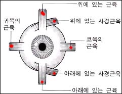
그림 13. 왼쪽 안구에 있는 근육, 한쪽 안구에 6개의 안구 근육이 정상적으로 붙어 있다.
Copyright ⓒ 2011 John Sangwon Lee, M.D., FAAP
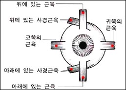
그림 14. 오른쪽에 있는 안구근육, 한쪽 안구에 6개의 안구 근육이 정상적으로 붙어 있다.
Copyright ⓒ 2011 John Sangwon Lee, M.D., FAAP
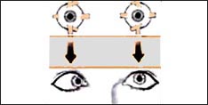
그림 15. 오른쪽 눈이 코 있는 쪽으로 쏠려 오른쪽 눈에 내 사시가 생겼다.
Copyright ⓒ 2011 John Sangwon Lee, M.D., FAAP
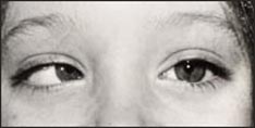
사진 16. 오른쪽 눈이 코 있는 쪽으로 쏠려 오른쪽 눈에 내사시가 생겼다
소스- 2011 Courtesey, Dr. Maynard B. Wheeler
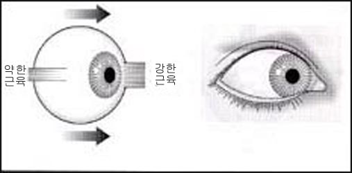
그림 17. 힘이 더 센 안구 근육이 붙어있는 쪽으로 안구가 쏠리어 사시가 생긴다. 내사시가 왼쪽 눈에 생겼다.
출처-Copyrightⓒ 2001 John Sangwon Lee,MD.FAAP
- 아래 그림 17과 같이, 귀 쪽에 붙은 안구 근육이 그 안구의 코 쪽에 붙은 안구 근육의 힘보다 더 약할 때는 오른쪽 안구가 코 쪽으로 끌어당겨져 안구가 코 쪽으로 쏠리게 되어 그 눈에 내 사시가 생긴다.
- 그림 15, 16과 같이, 왼쪽 눈의 6개의 안구 근육은 다 정상이다. 왼쪽 안구는 어느 한 쪽으로도 쏠리지 않고 정상적으로 움직일 수 있다. 그렇지만 오른쪽 안구의 6개 안구 근육 중 귀 쪽에 붙은 안구 근육의 힘이 그 안구의 코 쪽에 붙은 안구 근육보다 더 약하다. 따라서 오른쪽 안구가 코 쪽으로 쏠리게 된다. 이 때 오른쪽 안구에 내 사시가 나타나게 된다. 그래서 두 눈으로 한 개의 물체를 볼 때 두 안구가 조화 있게 정상적으로 움직일 수 없다.
- 12개의 안구 근육들 중 한 개나 그 이상 여러 개가 비정상적으로 더 약하게 되는 원인은 많다.
- 그 중 50%는 유전성이다. 드물게는 안구 근육에 분포된 제 3뇌신경, 제 4뇌 신경, 또는 제 6뇌신경의 마비나 신경이 손상 됐을 때 안구 근육에 마비현상이 생겨 사시가 생길 수 있다. 이런 이유로 생긴 사시를 마비성 사시라 한다.
- 안구 근육에 마비가 없이 생긴 사시를 비마비성 사시라고 한다.
- 드물게는 근무력증, 보툴리즘, 갑상성 기능 항진증 등으로 안구 근육 이상이 생겨 사시가 생길 수 있다.
신생아 사시 현상
- 사시의 증상은 나이, 사시의 정도와 종류, 원인 등에 따라 다르다.
- 신생아들이나 영아들의 눈에 실제로 사시가 없는 데 한쪽, 또는 양쪽 눈이 어느 한쪽 방향으로 일시적으로 쏠려 사시가 있는 것처럼 사시 현상이 생길 수 있다.
- 신생아들의 신체의 모든 근육들이 정상적으로 미약한 것처럼 신생아들의 안구 근육들도 미약하기 때문에 두 안구가 서로 조화 있게 잘 움직일 수 없는 것이 정상이다.
- 신생아들의 안구에 실제로 사시가 없는데도 일시적으로 사시 증상 징후가 나타날 수 있다. 이런 사시를 신생아 사시(Neonatal strabismus) 현상이라고 한다.
- 이와 같이 실제로는 사시가 없는데 사시가 있는 것처럼 보이던 신생아의 눈이 생후 3~4주 정도 지나면 양쪽 안구 근육들이 서로 조화 있게 잘 움직일 수 있게 되고 신생아 사시 현상이 잘 생기지 않는다.
- 신생아 사시 현상이 2~4 개월 이후 그냥 나타나거나 생후 2~4개월 이전이라도 친 부모형제 자매 중 사시가 있거나, 신생아의 눈에 다른 이상이 함께 있거나 걱정이 되면 단골 소아청소년 의사와 상담 한 후 안과전문의 확진을 받는 것이 적절하다(소스; Contemporary pediatrics, January, 2008. P18).
- 신생아기부터 사시가 있고, 사시의 증상 징후가 나타날 수 있다. 신생아에게 사시가 있다고 의심하면 의사에게 문의해야 한다.
- 부모도 반의사가 되어야 한다-제6권 신생아 성장 발육 융아 및 질병- 신생아가 사시가 있는 것 같은데요. 부모도 반의사가 되어야 한다- 제18권 소아청소년 이비인후과 질환-코 뿌리가 납작한 코 참조.
사시의 증상 징후
- 사시가 있는 눈으로 물체를 보려고 할 때 그 안구가 어느 한 쪽으로 쏠리고, 사시가 있는 쪽 눈 흰자위가 사시가 없는 정상 눈의 흰자위 보다 더 많이 보일 수 있다.
- 경미한 사시를 가지고 있을 때 힘이 약한 안구 근육에 무의식적으로 힘을 더 많이 주어 그 안구 근육이 정상 안구의 근육과 조화 있게 움직여 사시가 없는 것처럼 안구를 움직일 수 있다. 그 때문에 사시의 증상 징후가 잘 나타나지 않을 수 있다.
- 그러나 피곤하거나 아플 때는 안구 근육이 피로해지고 양쪽 안구의 전체 안구 근육의 힘이 야하게 된다.
- 이때 원래부터 약한 안구 근육이 있는 쪽 정 반대쪽에 붙어있는 정상 안구 근육이 있는 쪽으로 안구가 끌려가게 된다. 그 때 그 눈에 사시 현상이 현저하게 더 나타나게 된다.
- 어떤 물체를 정확하게 보기 위해서는 두 눈에 있는 12개의 안구 근육이 조화 있게 동시 움직여야 한다.
- 그렇지만 사시가 없는 정상적인 두 눈으로 어떤 물체를 볼 때도 두 눈이 조금씩 다른 각도에서 물체를 조금씩 달리 보고, 각 눈이 본 물체의 영상은 조금씩 다른 영상으로 뇌에 나타난다.
- 일반적으로 두 안구를 거쳐 들어온 두 영상을 뇌는 하나로 종합해서 그 물체를 선명한 한 개의 입체적 영상으로 만든다.
- 이렇게 두 정상 눈으로부터 들어온 한 개의 물체의 각 영상을 뇌 속에서 하나의 영상으로 종합해서 한 개의 입체적 영상으로 만드는 과정으로 그 물체의 입체감을 인지할 수 있고 그 물체가 얼마나 멀리 있는지 얼마나 가까이 떨어져 있는지 인지할 수 있다. (그림 18 참조).
- 심한 사시가 있을 때 한쪽 안구 근육, 또는 양쪽 안구 근육이 조화 있게 협력해서 움직이기가 어렵게 된다(그림 18 참조).
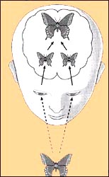
그림 18. 두 눈으로 한 개의 물체를 정상적으로 본 후 뇌 속에서는 두 눈으로 본 두 개의 영상을 한 개의 영상으로 종합한다.
출처-Copyrightⓒ 2001 John Sangwon Lee,MD.,FAAP
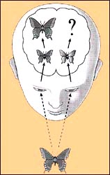
그림19. 사시가 있는 왼쪽 눈으로 본 한 개의 물체 영상은 희미하게 보일 수 있고 정상 눈으로 본 영상은 뚜렷하게 보인다.
출처-Copyrightⓒ 2001 John Sangwon Lee,MD.,FAAP
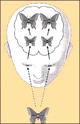
그림 20. 사시가 있는 눈으로 한 개의 물체를 보고 있는 데 뇌 속에 두 개의 영상이 동시에 만들어질 수 있다.
이와 같이 실제로는 하나인 물체가 두 개의 물체로 보이는 것을 복시라고 한다.
Copyright ⓒ 2011 John Sangwon Lee, M.D., FAAP
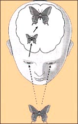
그림 21. 사시가 있는 왼쪽 눈이 본 물체는 뇌에서 정상적으로 인지되지 않을 수 있다.
이 때 왼쪽 눈의 시력이 약화될 수 있다. 이런 시력을 약시라고 한다. 더 심할 때는 그 쪽 눈의 시력이 상실될 수 있다. 이런 시력을 시약아라고 한다.
Copyright ⓒ 2011 John Sangwon Lee, M.D., FAAP
- 그래서 사시가 없는 정상 눈은 어떤 물체를 똑바로 볼 수 있지만 사시가 있는 눈은 코 있는 쪽이나 귀 있는 쪽, 눈 위쪽이나 눈 아래쪽으로 쏠려 물체의 입체감, 거리감을 제대로 인지 할 수 없다.
- 이 때 정상 눈이 인지한 물체의 영상은 뇌 속에서 뚜렷하게 인지될 수 있지만, 사시가 있는 눈이 본 물체의 영상은 뇌 속에서 희미하게 인지될 수 있다. 이런 경우 뇌는 두 영상을 한 개로 종합해서 하나의 영상으로 만들어 낼 수가 없다(그림 19 참조).
- 사시가 있을 때 두 눈으로 한 개의 물체를 보지만 한개 물체가 두 개로 보일 수 있다. 이것을 복시라고 한다 (그림 20 참조).
- 사시가 있는 눈으로 어떤 물체를 볼 때 뇌는 희미한 영상을 만들 수 있다. 따라서 그 물체는 희미하게 보일 수 있다.
- 사시가 있는 눈은 처음 얼마 동안은 복시 현상, 희미한 영상 등 문제를 해결해서 정확히 잘 보려고 노력하다가 이런 불편을 더 이상 극복할 수 없을 때는 사시가 있는 눈으로 더 이상 무엇을 보려고 노력하지 않고 주로 정상 눈으로만 보려고 한다.
- 더 시간이 지나가면, 사시가 있는 눈으로 더 이상 무엇이든지 보지 않고 사시 없는 정상 눈으로만 보게 된다.
- 이 때 사시가 있는 눈을 게으른 눈(Lazy eye)이라고 부른다.
- 이 게으른 눈으로 더 오랫동안 보지 않을 때 사시가 있는 눈의 시(각)신경의 기능이 점차로 퇴화될 수 있다. 결국 게으른 눈이 물체를 보고 그 물체의 영상의 거리감과 입체감을 뇌가 조금도 인지하지 못하게 된다.
- 이때 그쪽 눈의 시력이 점점 더 약화되어 결국은 그 눈으로 전혀 볼 수 없게 된다. 이런 결과로 생긴 약한 시력을 약시 또는 시약(Amblyopia)이라고 한다(그림 21 참조).
- 이런 약시를 가진 눈의 각막과 렌즈에는 큰 병변이나 변화가 생기지 않는 것이 보통이다.
- 사시로 인해 약시가 생긴 눈을 조기에 적절히 치료하지 않으면 그 눈은 영구히 완전히 멀게 된다.
- 시력 상실을 하게 하는 원인 중 가장 흔한 원인이 약시다.
- 이렇게 사시를 조기에 진단하여 치료하지 않으면 중대한 시력장애를 초래할 수 있다.
사시의 진단
- 병력, 증상 징후와 진찰소견을 종합해서 진단한다.
- 갓 태어나서부터 영유아기, 학령기, 사춘기를 거쳐 성인으로 성장될 때까지 모든 아이들이 소아청소년과나 클리닉에서 소아 건강검진을 주기적으로 받을 때 근시, 난시 또는 사시 등이 있나 눈에 다른 이상이 있나 진료 받는 것이 일반적이다.
- 소아청소년(0~18세)들이 소아 건강검진을 받을 때마다 눈과 시력검사를 기본적으로 받아야 한다.
- 일부 사시는 유전으로 생긴다.
- 친 부모 형제자매들 중 누구에게 사시가 있으면 다른 형제자매들에게도 사시가 있을 가능성이 많다.
- 친 부모형제 자매들 중 누구에게 사시가 있으면 그 집안 다른 아이들에게 사시가 있나 조심히 관찰해야 한다.
- 사시가 있다고 의심하면 소아 건강검진을 받을 때 부모는 의사에게 그 사실을 알려 주어야 한다.
- 아이에게 있는 심한 사시는 특별히 진찰하지 않고 그냥 눈을 보고 사시가 있는 것을 진단할 수 있다.
- 그러나 경미한 사시가 있을 때는 사시현상이 확연이 나타나지 않을 수 있다.
- 사시가 뚜렷하게 나타난다든지, 사시가 있다고 의심될 때 소아청소년과에서 머리부터 발끝까지 세밀한 진찰을 받고 의사의 소개를 받아 안과 전문의의 확진을 받고 적절한 치료를 받아야 한다.
- 신생아들이나 영유들에게도 사시 현상이 나타날 수 있지만 사시는 생후 2~3세 경에 더 현저히 나타나는 경우도 있다.
- 두 눈 사이에 있는 코 뿌리 뼈가 있는 비교(Nasal bridge)가 잘 발육되지 않아 코 뿌리가 납작하고, 더 넓고, 눈에 몽고인 추벽이 굵고 크게 생겼을 때 가성사시가 생길 수 있다.
- 실제로는 사시가 없는 눈에 사시가 있는 것 같이 보이는 경우를 가성사시, 또는 유사사시라고 한다.
- 이런 종류의 가성사시를 가진 아이가 점점 더 성장할 때 코 뿌리에 있는 뼈가 더 높아지고 커지면서 가성사시는 자연히 없어진다. 소아가정간호 백과 제18권 코 뿌리가 납작한 코 참조,
- 어느 때든지 시력에 이상이 있는 것 같으면 안과 전문의나 소아청소년과 전문의에게 문의해야 한다.
영유아의 시력검사
- 태어나서 2~3세가 될 때까지 시력검사표로 정확하고 자세한 시력을 검사할 수 없어도 다른 여러 가지 방법으로 영유아들의 시력을 어느 정도 정확하게 검사할 수 있고 대략 시력이 어떤지 가늠할 수 있다.
- 2~3개월 된 영아의 앞에다 작은 장난감을 갖다 놓으면 그 영아는 장난감을 보고 좋아하고 손으로 잡고 놀려고 한다. 영아가 장난감이 있는 쪽으로 얼굴을 돌리거나 장난감을 손으로 잡으려고 한다.
- 엄마를 보고 웃든지 무엇을 손으로 잡으려고 하는 것을 볼 수 있다.
- 이런 여러 종류의 눈의 발육 이정표로 영아들의 시력을 검사할 수 있다.
- 0~18세 소아청소년 건강검진을 받을 때마다 눈 검사와 시력검사를 받고,
- 3세나 그 이상이 되면 1년에 적어도 한 번 정도 눈 검사를 받고 시력검사표로 시력을 검사 받아야 한다.
- E 시력검사표로 시력검사를 받을 때 일부 유아들은 E 시력검사표를 잘 읽을 수 없다.
- 이럴 때는 E 시력검사표 대신 유치원 아이 그림 시력검사표 등으로 시력검사를 받을 수 있다.
- 어느 때든지 시력 이상이 있는 것 같으면 안과전문의나 소아청소년과에 문의해야 한다.
사시의 치료
- 사시가 있는 아이의 나이, 사시의 원인과 정도 등에 따라 치료한다.
- 사시가 있다고 진단 받았을 때 시력이 어느 정도인지 알아보고 시력에 따라 치료한다.
- 사시를 일으키는 약한 안구 근육을 튼튼하게 운동시켜서 사시를 치료할 수 있다.
- 사시가 없는 정상 눈에 안대를 대어 사시를 치료하거나
- 안경으로 치료 하거나,
- 사시가 있는 안구 근육을 수술 해 사시를 치료 할 수 있다.
- 사시를 언제, 어떻게 치료해야 가장 좋은지 마지막 결정은 안과전문의의 권장에 전적으로 맡겨야 한다.
- 일반적으로 사시가 있다는 것을 확실히 진단 받은 후 사시를 안경이나 안대, 또는 수술로 치료하든지,
- 앞서 설명한 여러 가지 사시의 치료 방법들 중 두세 가지 치료 방법을 함께 사용해서 치료해 줄 수 있다.
- 사시를 치료하는 방법에 대하여 다음 좀 더 자세히 구체적으로 설명한다.
안대( 눈 가리기)로 사시를 치료한다.
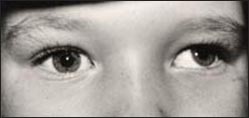
사진 22. 왼쪽 눈에 외사시가 있다
소스- Courtesey, Dr. Maynard B. Wheeler
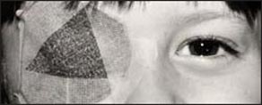
사진 23. 외사시를 안대로 치료할 수 있다
- 사시가 있는 눈에 약시가 항상 생기는 것은 아니다.
- 그렇지만 심한 사시를 제 때 적절히 치료하지 않으면 시력이 극도로 나빠져 약시가 생기고
- 그 약시를 적절히 치료하지 않으면 약시가 결국 실명으로 이어지고 눈이 먼다.
- 사시가 있는 눈에 약시가 생기지 않도록 예방적 치료를 하기 위해 건강하고 정상 눈에 안대를 얼마 동안 대어 사시가 있는 눈으로 물체를 보도록 치료한다.
- 안대로 사시를 치료해 주는 방법을 가장 많이 쓰는 사시치료이며 사시치료의 효과도 대단히 좋다.
- 그렇지만 이 방법으로 사시를 치료해도 사시가 완치되지 않는다.
- 사시를 안대로 치료할 때 정상적인 눈에 안대를 대고 사시 있는 눈으로만 얼마 동안 볼 수 있게 치료할 때 정상적인 눈에 안대를 대면 사시가 있는 눈은 온갖 힘을 다 해서 물체를 똑바로 보려고 노력을 한다.
- 그 때문에 사시로 약시가 이미 생겼던 눈이나 약시가 생기려고 하던 사시 있는 눈에 정상 시력이 다시 생길 수 있다.
- 의사의 지시에 따라 사시가 없는 정상적인 눈에 안대를 몇 주 동안 댔다가 다시 시력검사를 해보고, 필요하면, 그 후 의사의 지시대로 정상 눈에 안대를 계속 더 대어 사시를 치료한다. 그 다음 적절한 때 사시를 적절한 다른 치료를 할 수 있다.
- 아이가 어릴 때 사시를 안대로 치료하면 치료효과가 아주 좋다.
- 6~7세 이후 사시를 처음 진단 받았을 때 대부분의 사시가 있는 눈의 시력이 이미 상당히 나빠져 있기 때문에 그 눈에 약시가 심하게 생겨 있을 수 있다.
- 그래서 그 눈의 시력이 완전히 회복될 수 없는 때가 많다.
사시 안대 치료를 할 때 부모가 알아야 할 사항
- 두 눈 중 사시가 없고 잘 볼 수 있는 정상 눈에 안대를 대기 때문에 아이는 처음 얼마 동안 잘 볼 수 없고 사시가 있고 약시인 눈으로만 물체를 보아야 한다.
- 그래서 처음 안대 치료를 하는 동안 눈에 안대를 대고 다니기를 싫어한다.
- 그 외 다른 이유로 안대 대는 치료를 많이 싫어 할 수 있다. 그 때문에 어떤 아이는 성한 눈에 댄 안대를 자꾸 떼어버린다.
- 어떤 부모도 안대 대는 치료 효과를 잘 모르고 의사의 지시에 협조하지 않을 수 있다.
- 처음 치료를 시작한 얼마 동안 불편해도 그 환아 자녀 자신이 시력이 점점 회복되는 것을 느끼게 되고 안대를 대어 치료 하는 이유를 알게 되면 안대를 더 이상 떼 내지 않을 것이다.
- 때로는 안대 대어 치료를 절대로 하지 않으려는 아이도 가끔 있다.
- 이때는 안대에 그림을 그려 주는 등 다양한 방법을 써서 의사가 처방한 대로 안대 대는 치료를 한다.
- 온갖 노력을 다 해도 안대 대는 치료를 계속 할 수 없을 때는 안과 전문의와 상의해서 다른 방법으로 사시를 치료해야 한다.
- 의사의 지시 없이 부모 마음대로 정상 눈에 댄 안대를 떼서는 절대로 안 된다. 사시가 있는 눈에 생긴 약시가 회복될 기회를 영원히 잃어버리고, 그 눈이 평생 동안 멀 수 있기 때문이다.
사시를 안경으로 치료한다.
- 두 눈으로 어떤 물체를 볼 때, 두 눈이 동시 본 물체의 영상은 각막과 렌즈를 통해서 망막으로 보내진다.
- 이 때 눈은 렌즈를 크고 작게 적절히 변화시킨다.
- 즉 렌즈를 잘 조절(Accomodation)해서 본 물체의 영상을 망막으로 보낸다.
- 그렇지만 사시가 있는 눈이 본 물체의 영상은 본 그대로 망막으로 보내질 수 없는 때가 많다.
이런 사시는 안경으로 치료한다. - 사시를 안경으로 치료할 때도 사시를 근본적적으로 치료할 수 없다.

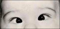
사진 24. 왼쪽 눈에 내사시가 있다
Courtesy, Dr. Maynard B. Wheeler 제공

사진 25. 사시를 안경으로 치료할 수 있다
눈 수술로 사시를 치료한다.
- 안구근육 수술로 사시를 치료하는 방법은 비교적 간단하다.
- 안구에 붙어 있는 안구 근육들 중 비정상적으로 더 강하고 더 짤막한 안구 근육이 있으면 그 안구 근육의 힘을 주리기 위해 그 강한 안구 근육을 느슨하게 하는 수술을 해 사시를 치료한다.
- 즉 좌우나 상하 대층 안구 근육 중 힘이 더 센 안구 근육의 정반대 쪽에 붙은 안구 근육의 힘과 거의 같은 힘을 갖게 안구 근육을 수술해 근육 힘을 동등 하게 조절 해 준다.
- 즉 힘이 약한 안구 근육을 수술해서 정상 안구 근육의 힘과 동등하게 만드는 수술치료를 한다.
- 사시를 수술로 치료해 줄 때 거의가 한 번 사시수술로 사시가 완치될 수 있지만, 어떤 때는 사시 수술을 두세 번 정도 해야 완치될 수 있다.
- 어떤 때는 사시가 있는 안구 근육을 수술해서 한쪽으로 쏠리는 안구를 정상 위치로 돌아가게 할 수 있지만 이미 생긴 약시는 수술치료로 완전히 회복시킬 수 없는 때도 있다.
- 수술로 사시를 치료해 준 이후에도 안대 대는 치료나 안경 사시치료를 동시 해 주어야 할 때도 있다.
- 또 사시수술 치료를 받은 이후 의사의 지시에 따라 눈과 시력을 주기적으로 검진 받아야 한다.
|
다음은 “사시의 원인, 종류, 치료”에 관한 인터넷 소아청소년 건강상담 질의응답의 예 입니다. |
Q&A. 사시의 원인, 종류, 치료에 관해
Q.
안녕하세요! 아이에게 무슨일이 생길때마다 이렇게 선생님께 문의 드리네요. 정말 고맙습니당. 다름이 아니오라 저희 아들 12개월된 남자아이 입니다. 쌍둥이라 제와절개로 36(1일)만에 태어났습니다. 쌍둥이치곤 아주 건강하세 낳았고요 2.6 2.8kg으로 낳았습니다. 산부인과 의사선생님께서도 아주 건강하고 칭찬도 해주셨는데. 여태껏 병원한번 안가고 예방접종외엔…건강했었는데… 제가 볼땐 잘 모르겠는데, 친정 엄마게서 아이가 눈이 한쪽바깥쪽으로 흩어진다고 계속 말씀을 하셔서 안과를 다녀왔습니당. 사실 이런건 믿기지 않아 제가 오히려 안믿고 싶어서 그런건지도 모르죠. 근데 “간헐성 사시” 인거 같다고 말씀을 하셨거든여?
수술을 받아야 한다고 말씀을 해주시고,,,제가 사는 곳이 경기도 안양인데 이곳 병원에서는 수술 할수있는 병원이 없다고 말씀을 하시더군요,,,아이가 쌍둥인데 어찌 한 아이만 이런지…
또 “간헐성 사시“가 유전인가여? 집안엔 사시가 업다고 들었는데.. 정말 답답합니다.
선생님 “간헐성사시“는 어떤 경우에 오는 것이며…어떻게 정말 수술밖에 치료방법이 없는것인지…알고 싶습니다. 부모되는것이 이렇게 어려운것인지 몰랐습니다.
A.
트윈님
안녕하세요. 또 질문해 주셔서 감사합니다. 좋은 질문입니다. 사정상 답변을 늦게 들여 죄송스럽습니다. 사실상 저는 소아청소년과 전문의이기 때문에 사시에 관한 의학지식이 안과 전문의에게 비하면 거의 없습니다.
사시의 원인 종류에 관한 정보가 이미 있지만 다음 다시 설명하겠습니다.
다음은 안과 참고서를 읽고 답변 드립니다.
불일치 사시(Incomitant strabismus)란 말은 있지만 간헐성(Intermittent)사시라는 병명을 찾을 수 없습니다.
혹시 교대성 사시(Alternating strabismus)를 의미하시는지요.
교대성 사시는 불안전 표현 율로 나타나는 체염색체 유전병입니다.
더 자세한 것은 안과에서 문의하시기 바랍니다.
다음 사시 정보를 참조하시기 바랍니다.
Cross-eye(Crossed eyes/Strabismus/Lazy eye/Squint/Eye deviation)-1
An overview of strabismus
• see strabismus-2
• A disease in which the coordination of the eyes does not work well because there is a deviation between the alignment of the visual axis of one eye and the alignment of the visual axis of the other eye is called strabismus (see Structure of the Eye).
• When looking at an object with both eyes, both eyes do not move in unison, one or both eyes are abnormally directed toward the nose, more toward the ears, downward or upward The more leaning is called strabismus, or squatting.
type of strabismus
• strabismus, in which the eyeball is directed inward (toward the nose),
• strabismus, in which the eyeball is tilted outward, is exostrabismus;
• Upper strabismus, in which the eyeball is tilted upward,
• strabismus, in which the eyeball is tilted downward is called downward strabismus.
• See strabismus classification
Several names for strabismus
• Strabismus is called Lazy eye, Crossed eyes, Cross-eyed, Squint, Strabismus, or Eye deviations.
• Also called ocular misalignment.
• Sashimi is also called four-fold or four-fold.
Early diagnosis and treatment of strabismus
• If strabismus is not properly treated in a timely manner, vision can become amblyopia and
• The amblyopia can lead to permanent blindness if not properly treated.
• Strabismus is one of the biggest causes of blindness.
• You may have only strabismus in your eyes.
• Sometimes strabismus can be caused by an underlying condition.
Causes of strabismus
• When the two eyeballs (eyeballs) do not work in harmony with each other, a deviation in the alignment of the visual axis occurs, resulting in strabismus.
• If the two eyes do not work harmoniously, the brain cannot create an image of the two objects seen by each eye into one image.
• Most strabismus diagnosed at or after birth are congenital. Such strabismus is called congenital strabismus.
• For the eyeball to move normally, the eyeball, orbit, eye muscle, cranial nerve, cerebrum, cerebellum, brainstem, vestibular nucleus, and oculomotor nucleus must function normally.
• The six eye muscles in each eyeball, eye muscle nerve development, and nerve function must have normal interactions, so that the eye’s visual axis can be arranged normally and one object can be viewed as one.
• Any abnormality in one or more of the eye muscles and ocular nerves listed above can cause strabismus.
Table 1. The following diseases or cases may cause strabismus.
표 1. 다음에 열거한 병이나 경우 사시가 생길 수 있다.
| 미숙아(Prematurity) | 태아 알코올 증후군(Fetal alcohol Syndrome) | 망막아세포종(Neuroblastoma) |
| 중추신경계 병(Diseases of central nervous system) | 두부 외상(Head trauma) | 뇌 외상(Brain trauma) |
| 눈 혈관종(Ocular hemangioma) | 두개 유합증( Craniosynostosis), | 아파트 증후군 (Apert syndrome) |
| 눈안 증후군(Noonann Syndrome) | 다운 증후군(Down syndrome) | 선천성 풍진 증후군(Congenital rubella Syndrome) |
| 뇌성 마비(Cerebral palsy) | 알라질 증후군( Alagille Syndrome | 그레이브스 병(Graves disease) |
| 근시(Nearsightedness/myopia) | 원시(Farsightedness/hyperopia) | 난시(Astigmatism) |
| 안검 종양(Palbral tumor) | 안구나 안와에 있는 병(Ocular or Orbital disease) | 당뇨병(Diabitis mellitus) |
| 눈 외상(Eye trauma) | 뇌 신경마비(Cranial nerve paralysis) | 알비니즘( 알비노현상/ 백피증/Albinism) |
| 근 무력증(Myasthenia gravis) | 라임병(Lyme disease) | 모바우스 증후군(Mobius syndrome) |
| 키아리 기형(키아리 기형, Chiari malformation) | 코넬리아 드랑게 증후군 (Cornelia de Lange Syndrome) | 엘러스-단로스증후군 ( Ehlers-Danlos syndrome) |
| 선상 모반 피지선 증후군(linear nevus sebaceous syndrome) | 프라더 윌리 증후군(Prader-Willi syndrome) | 루빈스타인-테이비 증후군(Rubinstein-Taybi syndrome) |
| 스미스-렘리-오피쯔 증후군(Smith-Lemli-Opitz syndrome) | 트리처 콜린스 증후군(Treacher Collins syndrome) | 터너 증후군이란(Turner syndrome) |
| 윌리엄스 증후군(Williams syndrome) | 색소 실조증(Incontinentia pigmenti) | 뇌량 무형성(agenesis of corpus callosum) |
References: Consultant for pediatricians, January 2008, p13 and “Internet This is an example of Q&A for children’s and adolescent health counseling” and others John Sangwon Lee Syndrome can cause strabismus and double vision. Copyright ⓒ 2011 John Sangwon Lee, M.D., FAAP
• As shown in Figure 13-14, 6 eye muscles are attached to one eyeball.
• A total of 12 eye muscles are attached to both eyes.
• When looking at an object with both eyes, it is normal for the 12 eye muscles to move in harmony with each other, moving both eyes in harmony to the direction of the object to be viewed, looking straight at the object and creating one image in the brain.
• Four eye muscles attached to the top and bottom, left and right of the eyeball, and 2 eye muscles attached to the top and bottom of the eyeball at an angle, a total of 6 eye muscles are attached to one eyeball.
• When one of the six eye muscles attached to one eyeball is abnormally weaker or stronger than the normal eye muscle strength,
• Strabismus occurs when the length of the superior or inferior ocular muscle is shorter or longer than the superior or inferior ocular muscle in the opposite direction of that eye (see figures 15, 16 below).
• For example, among the 6 eye muscles attached to one eyeball, when the muscle attached to the ear side of the eyeball is abnormally weak and the eye muscle strength to the nose side of the eyeball is normal, the eyeball will In this case, the nose is tilted towards the side, which results in strabismus (Fig. 16).
• Or, when the eye muscle attached to the ear of one eye is abnormally shorter but stronger than the eye muscle attached to the opposite side of the eye muscle, the eye moves toward the side of the ear with the abnormally shorter and stronger eye muscle. This can lead to exotropia in the eye.
Figure 13. Muscles in the left eye, 6 eye muscles are normally attached to one eye. Copyright ⓒ 2011 John Sangwon Lee, M.D., FAAP
Figure 14. The right eye muscle, 6 eye muscles are normally attached to one eye. Copyright ⓒ 2011 John Sangwon Lee, M.D., FAAP
Figure 15. My right eye is strabismus because my right eye is shifted towards the nose. Copyright ⓒ 2011 John Sangwon Lee, M.D., FAAP
Picture 16. My right eye is tilted towards the nose, resulting in esotropia in my right eye. Source – 2011 Courtesey, Dr. Maynard B. Wheeler
Figure 17. Strabismus occurs when the eyeball is tilted toward the side where the stronger eye muscles are attached. I have esotropia in my left eye. Source-Copyrightⓒ 2001 John Sangwon Lee, MD.FAAP
• As shown in Figure 17 below, when the eye muscle attached to the ear is weaker than the strength of the eye muscle attached to the nose of the eyeball, the right eyeball is pulled toward the nose and the eyeball is pulled toward the nose, resulting in strabismus in the eye.
• As shown in Figures 15 and 16, all 6 eye muscles of the left eye are normal. The left eye can move normally without tilting to either side. However, of the six eye muscles of the right eye, the strength of the eye muscle attached to the ear is weaker than the eye muscle attached to the nose of the eye. This causes the right eyeball to lean toward the nose. At this time, my strabismus appears in the right eye. Therefore, when looking at an object with both eyes, the two eyes cannot move normally in harmony.
• There are many causes of abnormal weakness in one or more of the 12 eye muscles.
• 50% of them are hereditary. Rarely, when the 3rd cranial nerve, 4th cranial nerve, or 6th cranial nerve distributed in the eye muscles is paralyzed or damaged, paralysis of the eye muscles may occur, resulting in strabismus. A strabismus caused by this reason is called paralytic strabismus.
• Strabismus that occurs without paralysis of the eye muscles is called non-paralytic strabismus.
• Rarely, myasthenia gravis, botulism, hyperthyroidism, etc. can cause ocular muscle abnormalities to cause strabismus.
Neonatal strabismus
• The symptoms of strabismus depend on the age, the degree and type of strabismus, and the cause.
• Newborns or infants who do not have strabismus in their eyes may have strabismus as if they had strabismus because one or both eyes were temporarily tilted in one direction.
• Just as all the muscles of a newborn’s body are normally weak, it is normal for the two eyes to move in harmony with each other because the eye muscles of newborns are also weak.
• Newborns may temporarily show signs of strabismus even when their eyes do not actually have strabismus. This type of strabismus is called neonatal strabismus.
• As such, when the eyes of a newborn baby, who appear to have strabismus even though they do not actually have strabismus, are around 3-4 weeks old, both eye muscles can move in harmony with each other and strabismus does not occur.
• If strabismus in newborns appears after 2~4 months of age, or even before 2~4 months of age, if any of your parents or siblings have strabismus, or if there are other abnormalities or concerns about the newborn’s eyes, consult with a regular pediatrician and see an ophthalmologist Confirmation by a specialist is appropriate (source; Contemporary pediatrics, January, 2008. P18).
• Strabismus is present in newborns, and symptoms of strabismus may appear. If you suspect that your newborn has strabismus, you should see your doctor.
• www.drleeepediatrics.com-Volume 6 Newborn Growth, Development, Childhood and Disease- It seems that the newborn has strabismus. Parents Should Be Anti-Doctors – See Volume 18 Children’s and Adolescent Otolaryngology Disorders – Nose with Flat Roots.
Symptoms, signs of strabismus
• When trying to see an object with the strabismus eye, the eyeball is tilted to one side, and the whites of the eyes with the strabismus may be seen more than the whites of the normal eyes without strabismus.
• When you have mild strabismus, you can unconsciously apply more force to the weak eye muscles so that the eye muscles move in harmony with the normal eye muscles to move the eye as if there is no strabismus. As a result, the symptoms of strabismus may not appear well.
• However, when you are tired or sick, the eye muscles become fatigued and the strength of the entire eye muscles in both eyes becomes weak.
• At this time, the eyeball is drawn to the side where the normal eye muscles are attached to the opposite side of the side where the original weak eye muscles are located. At that time, the strabismus phenomenon will appear in the eye significantly more.
• In order to see an object accurately, 12 eye muscles in both eyes must move in harmony and at the same time.
• However, even when looking at an object with two normal eyes without strabismus, the two eyes see the object from slightly different angles, and the image of the object viewed by each eye appears in the brain as slightly different images.
• In general, the brain synthesizes the two images that come in through the two eyes into one and creates a clear three-dimensional image of the object.
• In this way, the three-dimensional effect of the object can be recognized and how far and how close the object is by synthesizing each image of one object from the two normal eyes into one image in the brain to make one three-dimensional image. can be perceived (See Figure 18).
• In severe strabismus, it is difficult to move one or both eye muscles in a coordinated way (see Figure 18).
Figure 18. After seeing an object normally with both eyes, the brain combines the two images viewed with both eyes into one image. Source-Copyrightⓒ 2001 John Sangwon Lee, MD., FAAP
Figure 19. An image of a single object viewed with the strabismus left eye may be blurred, while images viewed with a normal eye are clearly visible. Source-Copyrightⓒ 2001 John Sangwon Lee, MD., FAAP
Figure 20. With a strabismus eye looking at an object, two images can be produced in the brain at the same time. In this way, when one object appears as two objects, it is called diplopia. Copyright ⓒ 2011 John Sangwon Lee, M.D., FAAP
Figure 21. Objects seen by the left eye with strabismus may not be recognized normally by the brain. In this case, vision in the left eye may be weakened. This vision is called amblyopia. In more severe cases, vision in that eye may be lost. This kind of vision is called a reagent child. Copyright ⓒ 2011 John Sangwon Lee, M.D., FAAP
• So, normal eyes without strabismus can see an object directly, but eyes with strabismus are focused on the nose or ears, above the eyes or under the eyes, so the three-dimensional effect and sense of distance cannot be recognized properly.
• At this time, the image of the object perceived by the normal eye can be clearly recognized in the brain, but the image of the object seen by the strabismus eye can be perceived faintly in the brain. In this case, the brain cannot combine the two images into one and create a single image (see Figure 19).
• When you have strabismus, you see one object with both eyes, but you may see one object as two. This is called double vision (see Figure 20).
• When looking at an object with a strabismus eye, the brain can produce a blurred image. Therefore, the object can be seen faintly.
• For the first time, the strabismus eye tries to see clearly by solving problems such as diplopia and blurred images, but when these inconveniences can no longer be overcome, the strabismus eye stops trying to see anything anymore and is usually normal. I’m just trying to see it with my own eyes.
• As time passes, the strabismus eye no longer sees anything and only the normal eye without strabismus.
• In this case, the eyes with strabismus are called lazy eyes.
• The function of the optic nerve of the strabismus eye may gradually deteriorate when the lazy eye does not look for a long time. Eventually, the lazy eye sees the object, and the brain does not recognize the distance and three-dimensional effect of the image of the object at all.
• At this time, the eyesight in that eye gradually weakens, and eventually the eye cannot see at all. The resulting weak vision is called amblyopia or amblyopia (see Figure 21).
• No major lesions or changes to the cornea and lens of the eye with such amblyopia are common. • Amblyopia caused by strabismus can result in permanent and complete blindness if not treated early and properly.
• Weakness is the most common cause of vision loss.
• If strabismus is not diagnosed and treated early, it can cause serious visual impairment.
Diagnosis of strabismus
• Diagnosis is made by combining medical history, symptoms, signs, and examination findings. • From birth to infancy, school age, puberty, and adulthood, when all children receive regular pediatric health check-ups at the pediatric department or clinic, check for myopia, astigmatism, or strabismus and other eye problems It is common to receive
• Whenever children and adolescents (0~18 years of age) undergo a pediatric health check-up, they should receive basic eye and vision examinations.
• Some lives are hereditary.
• If any of your biological parents’ siblings has strabismus, it is likely that other siblings will also have strabismus.
• If any of your biological parents or sisters have strabismus, you should carefully monitor other children in the household for strabismus.
• If you suspect that you have strabismus, you should tell your doctor when you have your child’s health checkup.
• Severe strabismus in a child can be diagnosed by looking at the eyes without special examination. • However, when there is mild strabismus, strabismus may not appear clearly.
• When strabismus is clearly visible or if strabismus is suspected, a detailed examination from head to toe in the Department of Pediatrics is recommended, a doctor’s referral is required, and an ophthalmologist confirms the diagnosis and receives appropriate treatment.
• Although strabismus may appear in newborns and infants, strabismus may appear more conspicuously around 2-3 years of age.
• Pseudostrabismus may occur when the nasal bridge between the two eyes is not well developed, so the nasal root is flat, wider, and the Mongolian wall is thick and large.
• A case in which an eye without strabismus appears to have strabismus is called pseudostrabismus or pseudostrabismus.
• As the child with this type of pseudostrabis grows, the bone at the root of the nose grows taller and larger and the pseudostrabis naturally disappears. See Encyclopedia of Pediatric and Home Nursing, Nose with Flat Nose, Volume 18,
• If at any time you suspect you have a vision problem, you should contact your eye care professional or pediatrician.
Eye examination for infants and toddlers
• Although it is not possible to accurately and detailed visual acuity tests from birth to 2 or 3 years of age, there are several other methods that can accurately test the visual acuity of infants and young children and give a rough estimate of their visual acuity.
• When a small toy is placed in front of a 2-3 month old infant, the infant will see and like the toy and will try to hold it and play with it. The infant turns her face toward the toy or tries to hold the toy in her hand. • You can see her smiling at her mother or trying to grab something with her hand.
• The developmental milestones of these different types of eyes can test infants’ vision.
• Children aged 0 to 18 years old receive an eye examination and vision examination at each health examination;
• Children aged 3 years and older should have their eyes checked at least once a year and have their eyes checked using the eye chart.
• When having an eye exam with the E eye chart Some infants cannot read the E eye chart well.
• In this case, you can take an eye exam using a preschool child’s picture eye exam table instead of the E eye exam sheet.
• If at any time you suspect you have a vision problem, you should contact your eye care professional or pediatrician.
Strabismus treatment
• Treatment depends on the age of the child with strabismus and the cause and severity of the strabismus.
• When you are diagnosed with strabismus, find out what your vision is and treat it according to your vision.
• Strabismus can be treated by strengthening the weak eye muscles that cause strabismus.
• Treat strabismus by applying an eye patch to a normal, non-strabismus eye.
• Treatment with glasses, or
• Strabismus can be treated by surgery on the eye muscles with strabismus.
• The final decision on when and how best to treat strabismus should be left entirely to the recommendation of an ophthalmologist.
• In general, after a definitive diagnosis that strabismus exists, strabismus is treated with eyeglasses, eye patches, or surgery;
• Among the various treatment methods for strabismus described above, two or three treatment methods can be used together for treatment.
• How to treat strabismus will be explained in more detail below.
Treat strabismus with an eye patch (covering the eyes).
Photo 22. Exotropia in the left eye Source – Courtesey, Dr. Maynard B. Wheeler
Photo 23. Exotropia can be treated with an eye patch Source – Courtesey, Dr. Maynard B. Wheeler
• Amblyopia does not always occur in strabismus eyes.
• However, if severe strabismus is not treated in a timely manner, vision can become extremely poor, resulting in amblyopia and
• If the amblyopia is not treated properly, it will eventually lead to blindness and blindness.
• As a preventative treatment to prevent amblyopia in the strabismus eye, a healthy, normal eye is patched for a period of time so that the strabismus eye can see objects.
• It is the most widely used method of treating strabismus with an eyepatch, and the effect of strabismus treatment is very good.
• Even if strabismus is treated with this method, strabismus will not be cured.
• When treating strabismus with an eye patch, putting an eye patch on the normal eye and treating only the strabismus eye with an eye patch When treating the normal eye with an eye patch, the strabismus eye tries with all its might to look directly at the object.
• As a result, normal vision may return to an eye that has already developed strabismus, or a strabismus eye that is about to develop amblyopia.
• As directed by your doctor, apply an eye patch to your normal, non-strabismus eye for a few weeks, then have your eyesight checked again, and if necessary, then continue to apply additional patches to your normal eye as directed by your doctor to treat strabismus. Then, when appropriate, other appropriate treatment for strabismus can be done.
• If you treat strabismus with an eye patch when your child is young, the therapeutic effect is very good. • When strabismus is first diagnosed after 6-7 years of age, most of the eyes with strabismus may have severe amblyopia because the eyesight has already deteriorated significantly.
• So there are many times when the eyesight cannot be fully restored.
What Parents Need to Know When Treating Strabismus Eyepatch
• Because the eye patch is placed on the normal, non-strabismic, well-seeing eye of the two eyes, the child must see objects only with the strabismus and low-vision eye for the first few years.
• That’s why I don’t like to wear an eye patch during my first eye patch treatment.
• For other reasons, you may be very reluctant to wear an eye patch. Because of this, some children keep removing the eye patch they put on their bright eyes.
• No parent may be unaware of the effectiveness of eye patch treatment and may not cooperate with a doctor’s instructions.
• Even if it is uncomfortable for some time after the first treatment, the child himself will feel that his or her eyesight is gradually improving, and when the child learns the reason for applying the eye patch, the eye patch will not be removed anymore.
• Sometimes, there are children who never want to be treated with an eye patch.
• In this case, using various methods such as drawing a picture on the eye patch, the treatment is performed as prescribed by the doctor.
• If, despite all your efforts, you are unable to continue with eye patch treatment, you should consult an ophthalmologist to treat strabismus in other ways.
• Never remove an eye patch from a normal eye without a doctor’s instruction. This is because the amblyopia in the strabismus eye permanently loses the chance to recover, and the eye can be blind for the rest of its life.
Treat strabismus with glasses.
• When looking at an object with both eyes, the image of the object viewed by both eyes is sent to the retina through the cornea and lens.
• At this time, the eye changes the lens large and small appropriately.
• In other words, the lens is well adjusted (Accommodation) and the image of the object is sent to the retina.
• However, the image of the object seen by the strabismus eye cannot be sent to the retina as it is seen.
This strabismus is treated with glasses.
• Even when strabismus is treated with glasses, strabismus cannot be cured fundamentally.
Picture 24. I have esotropia in my left eye Courtesy, Dr. Courtesy of Maynard B. Wheeler
Photo 25. Strabismus can be treated with glasses Courtesy, Dr. Courtesy of Maynard B. Wheeler Eye surgery to treat strabismus.
Picture 26. Both eyes have esotropia Courtesy, Dr. Courtesy of Maynard B. Wheeler
Photo 27. Some types of strabismus are treated with strabismus surgery. Copyright ⓒ 2011 John Sangwon Lee, M.D. FAAP
• The treatment of strabismus with eye muscle surgery is relatively simple.
• If there is an abnormally stronger and shorter eye muscle among the eye muscles attached to the eyeball, surgery to loosen the strong eye muscle to strengthen the eye muscle is performed to treat strabismus.
• In other words, the eye muscle is operated so that it has almost the same strength as that of the eye muscle attached to the opposite side of the eye muscle, which is stronger among the left and right or upper and lower ocular muscles, and equalizes the muscle strength.
• In other words, the weak eye muscles are operated on to make the strength of the normal eye muscles equal to the strength of the normal eye muscles.
• When strabismus is treated with surgery, almost all strabismus can be cured with one operation, but sometimes it can be cured by performing strabismus surgery two or three times.
• In some cases, the strabismus eye muscles can be operated to return the strabismus to its normal position, but in some cases the already formed amblyopia cannot be completely restored with surgical treatment.
• Even after surgical treatment of strabismus, it is sometimes necessary to treat strabismus with eye patch or glasses at the same time.
• After receiving treatment for strabismus surgery, you should have regular eye and vision checkups according to your doctor’s instructions.
The following is an example of Internet pediatric health counseling Q&A regarding “causes, types, and treatment of strabismus”.
Q&A.
About the causes, types, and treatment of strabismus Q. Good morning! Whenever something happens to my child, I ask the teacher like this. Thank you very much. It’s no different.
Our son is a 12-month-old boy. He was born at 36 (1 day) old with twins Raje and incision. I gave birth to twins who were very healthy and weighed 2.6 2.8 kg. The obstetrician-gynecologist was also very healthy and complimented me. I’ve never been to the hospital so far, except for vaccinations… I was healthy… I don’t know when I see it, but my mother kept telling me that my baby’s eyes were scattered to the outside, so I went to an ophthalmologist. Actually, I can’t believe this, so maybe it’s because I don’t want to believe it. But did you say that it sounds like “intermittent strabismus”? He told me that I had to have surgery, and he said that I live in Anyang, Gyeonggi Province, but there is no hospital that can perform surgery here. Is “intermittent strabismus” hereditary? I’ve heard that there is strabismus in the family… It’s really frustrating. Sir, I would like to know in what cases “intermittent strabismus” occurs… and how it really is the only treatment that is available…
I didn’t know being a parent could be so difficult.
A.
Twin Good morning. Thanks again for asking. That’s a good question. Sorry for the late reply due to circumstances. In fact, since I am a pediatrician, I have very little medical knowledge about strabismus compared to an ophthalmologist. There is already information about the types of causes of strabismus, but I’ll get back to you later. The following is an ophthalmology reference book and answer. There is a word called incomitant strabismus, but I can’t find the name of intermittent strabismus.
Do you mean Alternating strabismus? Alternate strabismus is a somatic chromosomal hereditary disease with an insecure expression rate. For more information, please contact your ophthalmologist. Please refer to the following isometric information.
출처 및 참조 문헌 Sources and references
- NelsonTextbook of Pediatrics 22ND Ed
- The Harriet Lane Handbook 22ND Ed
- Growth and development of the children
- Red Book 32nd Ed 2021-2024
- Neonatal Resuscitation, American Academy Pediatrics
- www.drleepediatrics.com 제1권 소아청소년 응급 의료
- www.drleepediatrics.com 제2권 소아청소년 예방
- www.drleepediatrics.com 제3권 소아청소년 성장 발육 육아
- www.drleepediatrics.com 제4권 모유,모유수유, 이유
- www.drleepediatrics.com 제5권 인공영양, 우유, 이유식, 비타민, 미네랄, 단백질, 탄수화물, 지방
- www.drleepediatrics.com 제6권 신생아 성장 발육 육아 질병
- www.drleepediatrics.com제7권 소아청소년 감염병
- www.drleepediatrics.com제8권 소아청소년 호흡기 질환
- www.drleepediatrics.com제9권 소아청소년 소화기 질환
- www.drleepediatrics.com제10권. 소아청소년 신장 비뇨 생식기 질환
- www.drleepediatrics.com제11권. 소아청소년 심장 혈관계 질환
- www.drleepediatrics.com제12권. 소아청소년 신경 정신 질환, 행동 수면 문제
- www.drleepediatrics.com제13권. 소아청소년 혈액, 림프, 종양 질환
- www.drleepediatrics.com제14권. 소아청소년 내분비, 유전, 염색체, 대사, 희귀병
- www.drleepediatrics.com제15권. 소아청소년 알레르기, 자가 면역질환
- www.drleepediatrics.com제16권. 소아청소년 정형외과 질환
- www.drleepediatrics.com제17권. 소아청소년 피부 질환
- www.drleepediatrics.com제18권. 소아청소년 이비인후(귀 코 인두 후두) 질환
- www.drleepediatrics.com제19권. 소아청소년 안과 (눈)질환
- www.drleepediatrics.com 제20권 소아청소년 이 (치아)질환
- www.drleepediatrics.com 제21권 소아청소년 가정 학교 간호
- www.drleepediatrics.com 제22권 아들 딸 이렇게 사랑해 키우세요
- www.drleepediatrics.com 제23권 사춘기 아이들의 성장 발육 질병
- www.drleepediatrics.com 제24권 소아청소년 성교육
- www.drleepediatrics.com 제25권 임신, 분만, 출산, 신생아 돌보기
- Red book 29th-31st edition 2021
- Nelson Text Book of Pediatrics 19th- 21st Edition
- The Johns Hopkins Hospital, The Harriet Lane Handbook, 22nd edition
- 응급환자관리 정담미디어
- Pediatric Nutritional Handbook American Academy of Pediatrics
- 소아가정간호백과–부모도 반의사가 되어야 한다, 이상원 저
- The pregnancy Bible. By Joan stone, MD. Keith Eddleman, MD
- Neonatology Jeffrey J. Pomerance, C. Joan Richardson
- Preparation for Birth. Beverly Savage and Dianna Smith
- 임신에서 신생아 돌보기까지. 이상원
- Breastfeeding. by Ruth Lawrence and Robert Lawrence
- Sources and references on Growth, Development, Cares, and Diseases of Newborn Infants
- Emergency Medical Service for Children, By Ross Lab. May 1989. p.10
- Emergency care, Harvey Grant and Robert Murray
- Emergency Care Transportation of Sick and Injured American Academy of Orthopaedic Surgeons
- Emergency Pediatrics A Guide to Ambulatory Care, Roger M. Barkin, Peter Rosen
- Quick Reference To Pediatric Emergencies, Delmer J. Pascoe, M.D., Moses Grossman, M.D. with 26 contributors
- Neonatal resuscitation Ameican academy of pediatrics
- Pediatric Nutritional Handbook American Academy of Pediatrics
- Pediatric Resuscitation Pediatric Clinics of North America, Stephen M. Schexnayder, M.D.
-
Pediatric Critical Care, Pediatric Clinics of North America, James P. Orlowski, M.D.
-
Preparation for Birth. Beverly Savage and Dianna Smith
-
Infectious disease of children, Saul Krugman, Samuel L Katz, Ann A.
-
Emergency Care Transportation of Sick and Injured American Academy of Orthopaedic Surgeons
-
Emergency Pediatrics A Guide to Ambulatory Care, Roger M. Barkin, Peter Rosen
-
Gray’s Anatomy
-
제19권 소아청소년 안과 질환 참조문헌 및 출처
- Habilitation of The handicapped Child, The Pediatric Clinics of North America, Robert H Haslam, MD.,
-
Pediatric Ophthalmology, The Pediatric Clinics of North America, Leonard B. Nelson, M.D.
-
Pediatric Ophthalmology, The Pediatric Clinics of North America, Lois J. Martyn, M.D.
-
Pediatric Ophthalmology, Edited by Robison D. Harley, M.D.
-
The Pediatric Clinics of North America, David Tunkel, MD., Kenneth MD Grundfast, MD
-
Atlas Pediatric Physical Diagnosis Frank A Oski
Copyright ⓒ 2014 John Sangwon Lee, MD., FAAP
“부모도 반의사가 되어야 한다”-내용은 여러분들의 의사로부터 얻은 정보와 진료를 대신할 수 없습니다.
“The information contained in this publication should not be used as a substitute for the medical care and advice of your doctor. There may be variations in treatment that your doctor may recommend based on individual facts and circumstances.
“Parental education is the best medicine.